Trizol is widely used because it combines tissue disruption with chemical separation in a single extraction workflow. For labs comparing RNA methods, its appeal is practical: it can recover RNA from many sample types while helping separate DNA and proteins. The chemistry relies on acid guanidinium thiocyanate and phenol-chloroform, so careful handling matters. Piotr Chomczynski and Nicoletta Sacchi described the approach in 1987: “A single-step method for RNA isolation by acid guanidinium thiocyanate-phenol-chloroform extraction is described.” That’s practical. But extraction quality depends on sound technique: complete homogenization, clean phase separation, and consistent pipetting help protect the aqueous RNA layer. A cloudy interface or careless transfer can carry contaminants into the sample. Small details matter. A brief pause at the interface can prevent accidental carryover, though it may cost a little yield. That trade-off is easy to miss.
Researchers should follow current product instructions, use suitable protective equipment, and assess RNA integrity and purity before downstream experiments. TRIzol is useful, not flawless. Phenol-based extraction may leave inhibitors, and yields can vary with tissue, sample input, and operator technique. For sensitive assays, validate the recovered RNA rather than assuming every preparation will perform equally. The method rewards care. And patience.
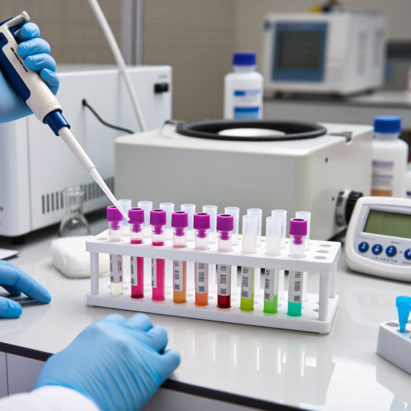
Phenol–guanidinium thiocyanate chemistry works by disrupting cells and rapidly denaturing proteins. Guanidinium thiocyanate also suppresses RNase activity, helping protect RNA from degradation. In the acidic phenol-based separation step, RNA remains mainly in the aqueous phase, while much of the DNA and protein partitions elsewhere. The method was described by Chomczynski and Sacchi in Analytical Biochemistry (1987; 162:156–159). The separation is tangible: after centrifugation, a clear upper layer sits above a cloudy interphase. Small details matter. Disturb that boundary, and genomic DNA may contaminate the RNA fraction.
The chemistry is powerful, but sample quality still depends on handling. Work quickly, keep tools RNase-free, and transfer only the aqueous layer. Still, not magic. Residual phenol or salt can affect downstream reactions, so assess purity and integrity rather than trusting appearance alone. The RNA Integrity Number (RIN) uses a 1–10 scale. ENCODE Consortium RNA-sequencing standards set a RIN value above 8 as a target for standard samples (ENCODE, 2012). That threshold is useful, not universal; degraded samples may still suit some applications. A clean-looking tube can mislead, and I would not treat one measurement as proof of success.
Sample loading sets the conditions for effective RNA release. A useful starting point is 1 mL of phenol-guanidinium lysis reagent per 50–100 mg of tissue. Smaller, softer pieces often disperse readily. Dense or fibrous tissue may need more thorough homogenization. Too much tissue for the available reagent can leave visible clumps and reduce contact with the lysis solution. That matters.
Weigh tissue before adding reagent, and trim it into small pieces with clean tools. Keep the sample cold during handling when practical, but avoid letting it thaw repeatedly. Add the measured reagent promptly, then homogenize until the mixture looks uniform, with no obvious chunks. A little stringiness can remain. It is not always easy to judge by appearance alone.
The 50–100 mg range is a guide, not a guarantee for every tissue type. Fatty, tough, or unusually dense samples may behave differently, so check the extraction protocol for the specific reagent and sample. If the lysate remains thick or uneven, reduce the tissue amount in a repeat preparation rather than assuming extra mixing will solve everything. I would also record tissue mass and reagent volume; those small notes make comparisons between samples more reliable.
During phase separation, add 0.2 mL chloroform for every 1 mL of phenol-guanidinium extraction reagent. This measured ratio helps separate RNA into the upper aqueous phase while proteins and DNA largely collect elsewhere. Cap the tube tightly, shake it firmly for about 15 seconds, then let it stand for 2–3 minutes at room temperature. Centrifuge according to the reagent protocol; many procedures use a chilled rotor. A distinct boundary should appear. It may look faint, so avoid rushing the transfer.
Tips: Use a calibrated pipette and a clean, RNase-free tube. After centrifugation, take the aqueous layer slowly, stopping before the cloudy interphase. Even a small amount of interphase carryover can affect RNA purity. If the boundary is unclear, pause and inspect the tube against a light background rather than guessing. Exact handling and centrifugation settings can vary by protocol, and the ratio alone cannot guarantee a clean sample. Record any changes; technique is not perfectly consistent from run to run.
A phenol-guanidinium extraction method helps separate RNA from proteins and other cellular material. The quality of the recovered RNA also depends on careful handling after phase separation. Once the aqueous phase has been mixed with the protocol-specified precipitation reagent, centrifugation collects RNA as a small pellet. It may be nearly invisible.
That detail matters.
Centrifuge at 12,000 × g for 15 minutes at 4°C when this setting matches your validated protocol. The low temperature helps limit degradation, while sufficient force allows the pellet to form. Use relative centrifugal force, not a guessed rpm value; rpm depends on rotor size.
After spinning, remove the supernatant gently without disturbing the pellet. A narrow pipette tip can help, but keep it away from the tube bottom. The pellet may sit along the side, not exactly at the center.
This step is easy to rush. If the supernatant looks cloudy or the pellet loosens, pause and check your protocol before proceeding.
Resuspend the pellet in nuclease-free water or the specified buffer, then assess concentration and purity before downstream work. Centrifugation settings vary among procedures, so consistency and careful observation matter.
A single tissue sample can contain several valuable molecular clues. An acid guanidinium-phenol extraction method can separate RNA, DNA, and protein from the same homogenized material. This is useful when sample amounts are limited or when comparing related molecular measurements. One tube, several fractions.
After phase separation, RNA is typically recovered from the upper aqueous layer. DNA may be collected from the interphase, while proteins remain in the organic phase or can be recovered from later washes. Careful pipetting matters: disturbing the boundary can carry contaminants into another fraction. Small details count.
This approach can make comparisons more consistent because each fraction begins with the same specimen. Still, recovery and quality vary with tissue type, sample freshness, and handling. Not always. A method that works well for cultured cells may perform differently with fibrous or lipid-rich tissue. Check each fraction’s purity and suitability before downstream assays, and follow the reagent’s safety instructions. It is also worth asking whether all three targets are needed; extra processing takes time and may reduce yield. Extraction is powerful, but it is not magic.
Phase separation can make RNA available from the aqueous phase, DNA from the interphase, and proteins from the organic phase of the same sample. This qualitative map shows primary collection fractions, not comparative yield; recovery depends on the sample and protocol.
Visit the official SA government portal www.sacoronavirus.co.za to stay informed about latest COVID-19 updates. Dismiss